Human Back Bones Anatomy / Human Spine By Mauriciokanno On Deviantart : The pelvis is composed of the two pelvic bones and the sacrum and coccyx (the pelvic bones are also known as the coxal, innominate, or hip bones) (fig.
Human Back Bones Anatomy / Human Spine By Mauriciokanno On Deviantart : The pelvis is composed of the two pelvic bones and the sacrum and coccyx (the pelvic bones are also known as the coxal, innominate, or hip bones) (fig.. The lumbar spine connects to the thoracic spine above and the hips below. The sacrum is a flat, triangular bone found in the lower back and wedged between the 2 hip bones. C1, c2, c3, c4, etc. The vertebral column is the defining characteristic of a vertebrate in which the notochord (a flexible rod of uniform composition) found in all chordates has been replaced by a segmented series of bone: The twelve thoracic vertebrae are numbered t1 to t12.
These bones are connected at the back with specialized joints. The range of motion in the thoracic spine is limited. Über 7 millionen englischsprachige bücher. Each are symmetrically paired on a right and left side. The vertebral column is a series of approximately 33 bones called vertebrae, which are separated by intervertebral discs.
Thoracic Anatomy Physiopedia from www.physio-pedia.com The shoulder joint is formed where the humerus (upper arm bone) fits into the scapula (shoulder blade), like a ball and. The human rib cage is made up of 12 paired rib bones; Posterior view of the lumbar spine and pelvis. The vertebral column of the lower back includes the five lumbar vertebrae, the sacrum, and the coccyx. Human body muscles human body organs human body parts human organ diagram body organs diagram anatomy organs anatomy bones heart anatomy body muscle anatomy. The column can be divided into five different regions, with each region characterised by a different vertebral structure. This framework consists of many individual bones and cartilages.there also are bands of fibrous connective tissue—the ligaments and the tendons—in intimate relationship with the parts of the skeleton. Bones are classified by their shape—as long, short, flat, and irregular.
The human rib cage is made up of 12 paired rib bones;
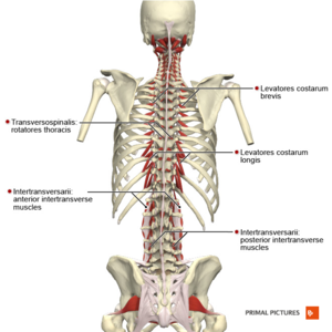
Bones, discs, and joints in your lower back your lower back contains 5 vertebral bones stacked above each other with intervertebral discs in between. Vertebrae, bones, joints, ligaments, muscles, muscular system, fascia, arteries, veins, nerves and various adjacent organs. A description of each of the vertebrae follows: Shop our carefully curated collection of high quality products today! Using this atlas of human anatomy of the spine and back. Some individuals may also have additional (i.e., supernumerary) cervical ribs or lumbar vertebrae. The sacrum is a flat, triangular bone found in the lower back and wedged between the 2 hip bones. It is situated towards the dorsal part of the torso. Über 7 millionen englischsprachige bücher. Human body muscles human body organs human body parts human organ diagram body organs diagram anatomy organs anatomy bones heart anatomy body muscle anatomy. The skeletal system consists of more than bones. Primarily, they are referred to as long or short. Touch device users, explore by touch or with.
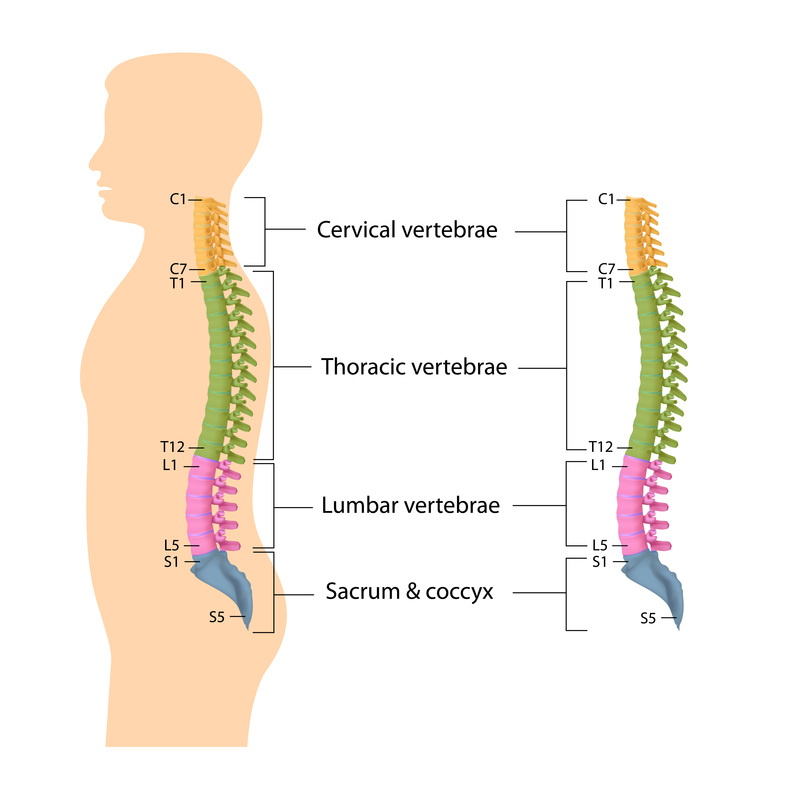
Cascade has served midwives and other healthcare professionals for over 40 years! The spine's four sections, from top to bottom, are the cervical (neck), thoracic (abdomen,) lumbar (lower back), and sacral (toward tailbone). The sacrum is a flat, triangular bone found in the lower back and wedged between the 2 hip bones. It comprises of a series of bones called the vertebrae of varying sizes extending from the skull to the small of the back. The range of motion in the thoracic spine is limited.
Information About Spine And Intervetebral Disc Anatomy from www.doneurosurgery.com The spine's four sections, from top to bottom, are the cervical (neck), thoracic (abdomen,) lumbar (lower back), and sacral (toward tailbone). There are 206 bones in the human skeleton, not including teeth and sesamoid bones (small bones found within cartilage): But look closer and you'll see even more structures. Posterior view of the lumbar spine and pelvis. There are small holes (foramen) along both sides of the sacrum that are left over when individual vertebrae fuse together. A description of each of the vertebrae follows: Human skeleton, the internal skeleton that serves as a framework for the body. The twelve thoracic vertebrae are numbered t1 to t12.
Human body anatomy female female anatomy muscle shoulder blade pain anatomy back muscles bones man female anatomy body muscles in a body female anatomy muscole shoulder concept muscular sysyem.
The cervical spine consists of 7 vertebra that are numbered 1 through 7 from top to bottom i.e. The pelvis is composed of the two pelvic bones and the sacrum and coccyx (the pelvic bones are also known as the coxal, innominate, or hip bones) (fig. Read on to get 10 key facts about the human skeleton. Cascade has served midwives and other healthcare professionals for over 40 years! C1, c2, c3, c4, etc. Human bones diagram 12 photos of the human bones diagram human anatomy diagram back view organs, human anatomy diagram diaphragm, human anatomy diagram of ear, human anatomy torso diagram, human skeleton diagram with labels, bone, human anatomy diagram back view organs, human anatomy diagram diaphragm, human anatomy diagram of ear, human. This includes the head, facial, hyoid, auditory, trunk, ribs, and sternum. While in the thoracic and lumbar spine, the anatomy of the vertebrae is relatively consistent between each vertebra, cervical spine anatomy is quite variable. The vertebral column is the defining characteristic of a vertebrate in which the notochord (a flexible rod of uniform composition) found in all chordates has been replaced by a segmented series of bone: The lumbar spine is composed of five vertebrae, named l1 to l5 from superior to inferior. A ridge across the front (anterior) portion of the s1 vertebra is called the sacral promontory. See human back anatomy stock video clips. When autocomplete results are available use up and down arrows to review and enter to select.
The bones provide a structural framework and protection to the soft organs. Vertebrae, bones, joints, ligaments, muscles, muscular system, fascia, arteries, veins, nerves and various adjacent organs. The vertebral column of the lower back includes the five lumbar vertebrae, the sacrum, and the coccyx. The most common variations include sutural (wormian) bones, which are located along the sutural lines on the back of the skull, and sesamoid bones which develop within some tendons, mainly in the hands and feet. Vertebrae separated by intervertebral discs.
Human Spine Disorders Anatomical Chart Anatomical Chart Company Amazon De Books from m.media-amazon.com The bones provide a structural framework and protection to the soft organs. Human bones diagram 12 photos of the human bones diagram human anatomy diagram back view organs, human anatomy diagram diaphragm, human anatomy diagram of ear, human anatomy torso diagram, human skeleton diagram with labels, bone, human anatomy diagram back view organs, human anatomy diagram diaphragm, human anatomy diagram of ear, human. Each are symmetrically paired on a right and left side. Primarily, they are referred to as long or short. Bone tissue anatomy and physiology 12 photos of the bone tissue anatomy and physiology anatomy and physiology bone tissue test, anatomy and physiology chapter 6 skeletal system bone tissue, anatomy and physiology osseous tissue and bone structure quiz, bone anatomy and physiology ppt, bones anatomy and physiology test, bone, anatomy and. From media.istockphoto.com however, as a child grows, some of the bones fuse together. The pelvis is composed of the two pelvic bones and the sacrum and coccyx (the pelvic bones are also known as the coxal, innominate, or hip bones) (fig. Vertebrae separated by intervertebral discs.
See human back anatomy stock video clips.
On anatomical parts the user can choose to display the various structures in colored illustrations of the anatomy of the back and spine: The sacrum is an irregular (sphenoid) bone that makes up the back (posterior) third of the pelvic girdle. The sacrum is a flat, triangular bone found in the lower back and wedged between the 2 hip bones. Vertebrae, bones, joints, ligaments, muscles, muscular system, fascia, arteries, veins, nerves and various adjacent organs. The lumbar spine connects to the thoracic spine above and the hips below. The human rib cage is made up of 12 paired rib bones; This framework consists of many individual bones and cartilages.there also are bands of fibrous connective tissue—the ligaments and the tendons—in intimate relationship with the parts of the skeleton. A ridge across the front (anterior) portion of the s1 vertebra is called the sacral promontory. Über 7 millionen englischsprachige bücher. Human body anatomy female female anatomy muscle shoulder blade pain anatomy back muscles bones man female anatomy body muscles in a body female anatomy muscole shoulder concept muscular sysyem. While in the thoracic and lumbar spine, the anatomy of the vertebrae is relatively consistent between each vertebra, cervical spine anatomy is quite variable. There are small holes (foramen) along both sides of the sacrum that are left over when individual vertebrae fuse together. The skeletal system includes over 200 bones, cartilage, and ligaments.


0 Komentar